
The upper front jaw is one of the least forgiving implant sites. The nasopalatine canal sits directly behind the central incisors, and the buccal bone in front of the roots is often thin. A panoramic or periapical image cannot show either reliably. A CBCT can, and the report should state both explicitly.
Placing an implant in the aesthetic zone looks simple on a flat image: there is bone, there is space, and the neighboring teeth give a guide. The difficulty is that the two structures that most often decide the outcome, the nasopalatine canal and the buccal bone plate, lie in the one plane a flat image cannot see, which is front to back. Why 3D imaging is considered essential for implant planning covers the wider case for the scan.
A surgeon who looks at a clean periapical before an upper incisor implant is looking at the one view that hides the two things most likely to go wrong. Below: what those two structures are, how often they turn out to be a problem according to published CBCT series, and what a scan report has to say about each. It describes hazards and questions to ask, not a measurement protocol.
Why the Anterior Maxilla Is a High-Risk Site
Several things combine in this small region.
Thin bone in the wrong direction. The ridge is often adequate in height and still narrow from front to back, with a labial plate that can be very thin.
A large canal in the midline. The nasopalatine canal runs from the nasal floor to the palate, close to the roots of the central incisors. Its lower-jaw counterpart has variants of its own, covered in mandibular canal and mental foramen variations.
Visible consequences. A failure here is seen every time the patient smiles, through recession, grey show-through or a lost papilla.
Immediate placement. Many implants in this zone are placed into a fresh extraction socket, where the walls are already compromised.
The Nasopalatine (Incisive) Canal
The nasopalatine canal, also called the incisive canal, carries the nasopalatine nerve and a small artery from the nose to the palate. It opens in the midline behind the upper central incisors at the incisive foramen. It is a normal structure, and it is variable.
What varies
Diameter. In one CBCT study of 198 scans from an Iranian population, the canal measured on average about 3.5 mm across at the incisive foramen, narrowed to about 2.4 mm mid-canal and widened to about 3.7 mm at the nasal opening. It was wider in edentulous patients (about 4.1 mm) than in dentate patients (about 3.5 mm) [2]. Other populations differ.
Number of openings. In the same series about 82% of canals had a single palatal opening, and about 18% had two or three [2].
Shape. The cylindrical form was most common (about 58%), with funnel, hourglass, banana and Y-shaped forms making up the rest [2].
Length. Mean canal height was about 12.8 mm, with a wide range of roughly 5 to 21 mm [2].
Direction and nasal opening. Both affect how far an implant can safely extend upward, and neither is shown on a flat image.
A CBCT series of 100 consecutive patients examined the canal together with the buccal plate in front of it, because these two decide the plan together [1].
A normal canal that becomes a problem
A canal does not have to be abnormal to cause trouble. Because it lies directly behind the roots of the central incisors, an implant placed in or beside a central incisor socket can come into contact with it. If the canal is wide, the available bone may be little more than the width of the socket.
When it is enlarged by disease
A nasopalatine duct cyst can widen the canal and thin the bone around it. These are among the more common jaw cysts, often found by chance. A widened canal in a planning scan is a reason to ask whether it is simply a large normal canal or a lesion, because the answer changes the plan. How cysts and tumors are recognized on CBCT covers the wider principles, and a case in which a lesion was not what it first appeared shows what can go wrong when imaging is read too quickly.
What Goes Wrong When an Implant Meets the Canal
Failed integration. Soft tissue in the canal means no bone contact along part of the implant, and the implant may fail to stabilize.
Altered sensation. Pressure on the nasopalatine nerve can cause numbness or tingling of the front of the palate.
Bleeding. The small artery in the canal may bleed during drilling.
Late failure or infection. An implant that sits partly in the canal may not remain healthy.
Nasal complications. Over-extension can perforate the nasal floor.
Not every contact is a failure. Some clinicians deliberately manage the canal, for example by modifying the plan or the technique, which is a surgical decision that depends on knowing exactly what is there.
Buccal Bone in the Aesthetic Zone
The buccal, or labial plate is the thin wall of bone in front of the roots of the upper incisors, and it is thinner than most clinicians expect. A systematic review and meta-analysis of CBCT studies (17 studies, more than 9,000 teeth) found a mean thickness of roughly 0.75 to 1.05 mm for the anterior teeth. About 70% of central incisors, 65% of lateral incisors, and 55% of canines had a wall thinner than 1 mm, and about 30% of central incisors and 35% of lateral incisors had a wall thinner than 0.5 mm [3]. Heterogeneity between studies was high, so treat these as an indication of the pattern and not as a prediction for one patient. Earlier CBCT work described the same pattern [4].
That matters because the plate is supported by the tooth root. When the tooth is removed, the plate loses the blood supply and function that it depends upon, and it tends to resorb. The result can be recession of the gum, an exposed implant collar, or a collapse of the contour that no crown can hide later.
What the plate depends on
Thickness. Thicker plates resorb less, and thin ones can be lost partly or fully after extraction.
Integrity. A fenestration or a dehiscence in the plate, which may already exist before surgery, weakens the site.
Root angulation. The root often sits at an angle to the ridge, leaning forward. An implant placed along the ridge may lie too close to the plate or emerge through it.
Labial concavity. A concavity below the apex of the incisors changes where the implant can be placed without perforating.
Gum phenotype. Thin gum tissue shows any change in the underlying bone.
Immediate placement and the buccal gap
Placing an implant into a fresh socket is attractive, and in the anterior maxilla it is also the highest-stakes version of the procedure. The usual surgical principle is to keep the implant towards the palatal wall of the socket, leaving a gap on the buccal side. Many clinicians aim for a gap of around 2 mm, and a plate thickness of at least 1 to 2 mm after placement; check these against your own protocol. Whether those margins can be achieved depends entirely on the real thickness, position and angulation of the plate, which are exactly the measures a flat image cannot give.
Does a Thin Plate Mean an Immediate Implant Will Fail?
No. A thin plate raises the risk of recession and contour loss, and it does not make failure inevitable. What it does is narrow the margin for error. The surgeon who knows the plate is thin can choose a staged approach, graft the buccal gap, place the implant more palatal, use a thicker-phenotype soft tissue graft or accept a compromise on the final contour. The surgeon who does not know, is making the same decisions blind-folded. The scan changes the decision from a guess to a choice, and that is its real value here.
Why 2D Images Under-Report Both
| Structure | What a periapical or OPG shows | What it cannot show |
|---|---|---|
| Nasopalatine canal | A dark vertical band between the incisor roots, if the angle is right | Its width from front to back, its true shape, branches, and its distance from the socket |
| Buccal plate | Not reliable. The plate lies edge-on to the beam and is superimposed on the root | Its thickness, any fenestration or dehiscence, and its contour |
| Root angulation | The root outline in two dimensions | Its direction relative to the ridge, and the likely emergence point of an implant |
| Labial concavity | Not visible | Its depth and position |
| Nasal floor and canal opening | A faint outline at best | Their height above the planned implant apex |
The underlying limit is that a periapical or panoramic image compresses the jaw into a plane. Everything lying in front of or behind a structure is stacked on top of it. The two features that decide anterior implants are both measured from front to back, and they are the ones most hidden. The imaging mistakes behind implant failures, shows where planning could go wrong.
What a CBCT Adds, and Its Limits
A CBCT shows the area in cross-section, so the canal, the plate, and the root can be seen from the side. This allows the canal's width and branches, the plate's thickness, any fenestration or dehiscence, the root's angulation and the concavity below it to be examined directly.
It is not perfect.
Thin structures are hard to see. A very thin buccal plate, below the resolution of the scan, may appear absent when it is present. A report that says "plate absent" should say whether it is truly absent or below the limit of detection.
Artefact. Metal restorations in neighboring teeth, or a crown on the tooth being replaced, can obscure the plate.
Resolution depends on the settings. A larger field and larger voxels give less detail for thin walls. How to choose the field of view explains the trade-off.
Soft tissue is not shown. Gum thickness and phenotype are judged clinically.
Measurements carry uncertainty. Estimates can vary by a fraction of a millimeter or more, which matters when the plate is only a millimeter thick.
What the Report Should State
For an anterior maxillary implant plan, a thorough report says more than "adequate bone." Ask for the following:
The size and shape of the nasopalatine canal, whether it has more than one channel and where it lies relative to the planned site.
Any widening or lesion associated with the canal.
The buccal plate thickness at the planned site, and whether any fenestration or dehiscence is present.
The angulation of the root relative to the alveolar ridge.
The presence of a labial concavity.
The height of the bone to the nasal floor.
The limits of the scan, such as artefact or a field that cuts off the site.
If the report cannot state these, the request may not have specified them, or the field of view may not have covered them. A section-by-section guide to CBCT reports shows what to expect, and a guide for referring Dentists covers writing a request that gets them.
Reading It Alongside the Mouth
A scan describes bone. The patient's smile, gum thickness, lip line and the condition of the neighboring teeth decide how much bone loss the site can tolerate. A patient with a high smile line and thin tissue has little room for any collapse, and the plan should account for it. A patient with a low smile line and thick tissue is more forgiving. The scan and the clinical picture answer different halves of one question.
Planning Choices That Follow the Findings
A wide canal near the socket. The plan might shift the implant position, change its diameter, or consider managing the canal as part of the procedure.
A very thin or missing plate. The surgeon may choose a staged approach, grafting before or at placement, or a technique that protects the plate.
A forward-leaning root. The implant axis and the restoration are planned together, often with a screw-retained crown and sometimes an angled component.
A concavity. The implant depth and angle are chosen to avoid perforating the labial wall.
A lesion in the canal. The plan waits for assessment and treatment of the lesion.
These are examples of how the findings steer judgement. Each decision belongs to the surgeon and depends on skills beyond this article.
A Short Illustrative Scenario
Consider an imagined patient with a fractured upper central incisor that cannot be saved. A periapical image shows adequate root length and no obvious problem. The surgeon plans an immediate implant. A CBCT before surgery, however, shows a wide nasopalatine canal that sits directly behind the socket, and a buccal plate that is thin and has a small fenestration near the apex. The plan changes: the implant position is adjusted, the buccal gap is managed and grafting is planned. Without the scan, the implant might have entered the canal or pushed through a plate that could not support it.
This is a composite illustration and not a record of a real patient. The pattern, however, is a familiar one.
Where Sinus and Nasal Structures Come In
In the anterior maxilla, the nasal floor and the nasopalatine canal opening limit implant length. Where the plan extends to the premolar region, the maxillary sinus becomes relevant and the imaging checklist for bone grafting and sinus lifts covers it. What Dentists miss in the maxillary sinus without CBCT is a useful companion for planning in the posterior maxilla.
Immediate or Delayed Placement: How the Scan Informs the Choice
The scan does not make the decision, but it frames it. A thick, intact plate, a canal well behind the socket and a root that lies close to the ridge axis make immediate placement more predictable. A thin or defective plate, a wide canal against the socket or a strongly forward-leaning root push towards a staged approach, with grafting or a delay to allow healing. In many anterior cases the honest answer is that immediate placement is possible but leaves little margin, and the decision rests on the surgeon's experience and the patient's expectations.
It is worth telling the patient this plainly. A smile that is visible every day is the place where an optimistic plan produces the most disappointing result. Explaining that the scan found a thin plate, and that the plan has been adjusted for it, builds more trust than a promise that everything will look perfect.
Questions the Patient May Ask, and Honest Answers
"Why do I need a 3D scan when my X-ray looked fine?" Because the structures that help to decide the implant, site lie in the front-to-back direction, and are not seen a flat X-ray.
"Can the implant touch the canal?" Sometimes the plan manages it deliberately, and sometimes it is avoided. Either way, the surgeon should know exactly where the canal is.
"Will my gum recede?" Thin bone and thin gum raise the risk, and the plan can reduce it but not remove it entirely.
"Can everything be done in one visit?" Sometimes. The scan helps decide whether that is wise.
A Note on Training
Recognizing canal variants, thin plates and root angulation on a CBCT is a skill built by working through volumes in several planes, many times. A structured course in implant-focused CBCT reading, such as CBCT Implantology, is one route, and a conversation with the reporting Radiologist about each case is another.
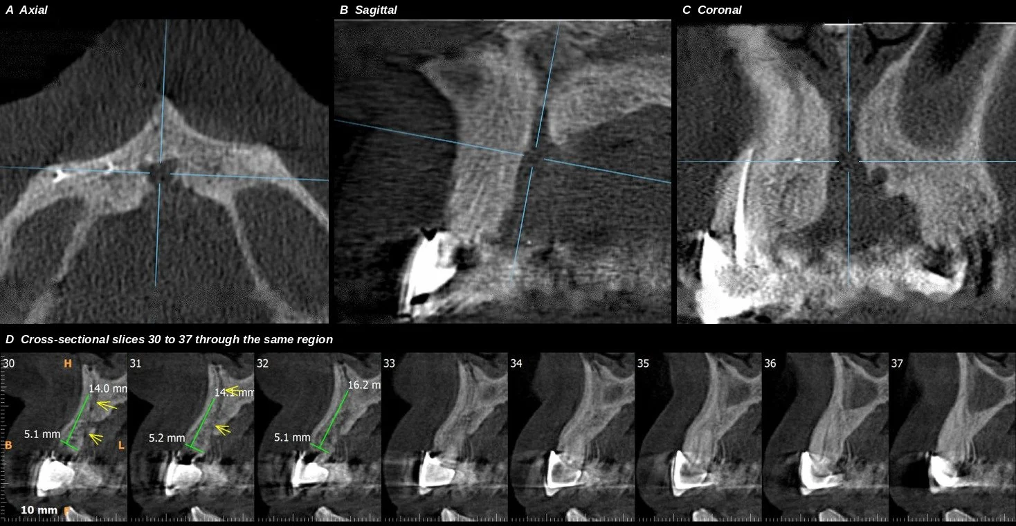
Figure. De-identified CBCT of the upper front jaw. A to C: axial, sagittal and coronal views, with the crosshairs on a prominent nasopalatine canal. D: cross-sectional slices through the same region, with the annotations from the original scan report.
A patient, presented for a CBCT scan to evaluate available bone in the anterior maxilla for planning dental implants. Patient was using bridge since 15 yrs but now wants to go for more advanced treatment.. The bone volume appeared adequate on the previous Panoramic (OPG) X-ray; however, the CBCT sections revealed a prominent nasopalatine canal and asymmetric remodeling of the residual alveolar ridge.
A Checklist Before the Implant Appointment
Is the nasopalatine canal shown, with its width, shape, and distance from the socket?
Is the buccal plate measured and described at the planned site?
Is there a fenestration, dehiscence or concavity?
What is the root angulation relative to the ridge?
How far is the nasal floor above the planned implant?
Is the field of view and the voxel size suitable for thin structures?
Does the plan allow for what the scan could not show?
Frequently Asked Questions
-
A canal in the midline of the upper jaw, behind the central incisors, carrying a nerve and a small artery from the nose to the palate. It is a normal structure that varies in size and shape.
-
An implant placed in or against the canal can cause failed integration, altered sensation in the front of the palate, bleeding, or other problems. Planning with a 3D scan helps avoid or manage it.
-
The labial plate in front of the incisor roots is naturally thin, often about a millimeter or less, and it depends on the tooth root for support. It can undergo resorption after extraction.
-
No. The plate lies edge-on to the beam, so thickness cannot be judged. A CBCT shows it in cross-section.
-
Clinicians generally want as much as possible, and many aim for at least 1 to 2 mm after placement along with a buccal gap. The numbers vary by technique and authority, so the surgeon's protocol applies.
-
The canal's width, shape and branches, any lesion, the buccal plate thickness and integrity, root angulation, any concavity, the height to the nasal floor and any limits of the scan.
Related Reading
Questions about a specific scan or report? The contact page lists how to reach the team.
